

According to official data, more than 2 million helminth infections have been recorded in Russia. These numbers are constantly increasing, judging by the number of pharmaceuticals sold annually, worms in the feces of a child or an adult are more common.
Today there are about 350 types of parasites, in our country there are 70. It is not always possible to determine that the baby is infected immediately, the worms can live for some time, practically not making themselves felt in the meantime, destroying the body, eating useful substances from food ...
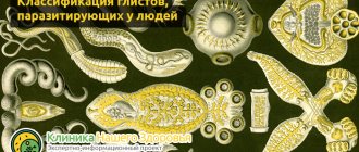
Types of worms in children in feces
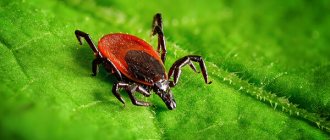
Children are more likely to be at risk of contracting helminths. They study the world around them every day, trying to taste each object not only by touch, but also by taste.
Parents can learn about the infection only at the acute stage of the disease. If ignored, it becomes chronic.
Without adequate and timely medical care, the invasion will not only weaken the immune system, but will negatively affect the functioning of the whole organism.
Usually, children become infected with pinworms - they cause the development of enterobiasis. This type of parasite settles in the small intestine and lays many eggs there in a couple of weeks of its existence.
Pinworms
Also, a child can become infected with roundworms - these worms can grow up to 40 cm in length. They colonize the intestines, but do more damage than pinworms. There are other types of parasites, but infestation is less common.
Ascaris


Fluke class flatworms
In medicine, this type of parasite is called fluke or fluke. In a child, these worms cause trematodosis disease with the localization of pathogens in the liver, lungs and circulatory system. Flukes are small in size from 1–2 mm to several centimeters.
Hepatic fluke
This is a zoonotic helminth that lives in the ducts of the liver and causes the parasitic disease fascioliasis in children. A sexually mature individual reaches 5 cm in length and has a leaf-shaped body. The abdominal and oral parts of the parasite have one sucker each.
The testes are located on the front of the body. In the middle, under the abdominal sucker, there is a segmented uterus and an ovary. On the lateral part of the liver fluke, you can see the vitellines, which serve as a source of nutrition for the embryos.
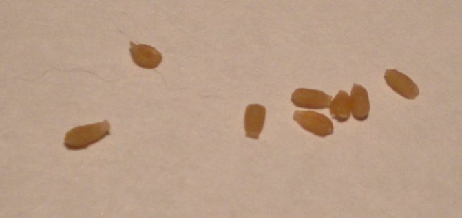
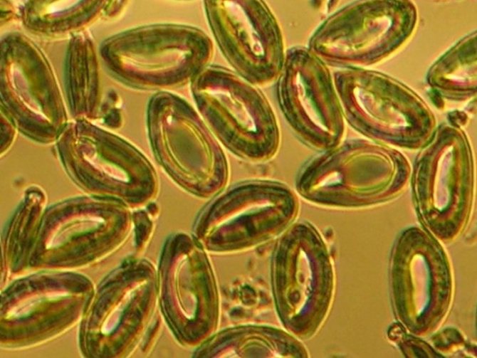
It will not be possible to see the eggs of this type of worm in the feces without a microscope. Their dimensions are less than a millimeter in length. A characteristic cap is located at one end. Eggs are usually yellowish or light brown in color.
Children are not infected with fascioliasis by eating infected intermediate host meat. You can get sick only when contaminated water enters the mouth, where fully mature larvae (adolescaria) live.
Feline fluke
The parasite is called Siberian fluke, cat fluke or Siberian fluke. The worm got its name in connection with the first detection of individuals of this type in the body of cats. Flukes are the causative agents of opisthorchiasis, a dangerous disease for a child, which affects the liver.
The worms grow up to 13 mm in length and are leaf-like, slightly tapered in front.At the top of the body there is a sucker, from which the pharynx and two intestinal loops, closed and extended along the entire parasite, stretch. In the middle of the worm there is an abdominal sucker, followed by a uterus with ramifications and an ovary. Two testicles can be seen behind the body. The structure of the parasite also gave the name to the disease, from the Greek Opisthorchis means the testis behind.
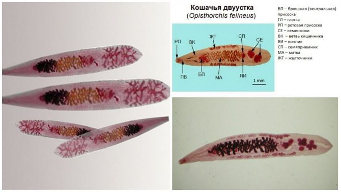
Feline fluke
Microscopic examination can show the eggs of these worms in baby feces.
Up to 800 fluke larvae can be found in 2 grams of baby feces. They are microscopic in size and resemble a lemon in color and shape. The upper part of the egg has a cap and is narrower than the lower pole. Inside the cocoons, the contents of a heterogeneous structure are in the form of small grains.
Opisthorchiasis is diagnosed only in children who eat fish products, therefore, the disease is not common in infants.
Another parasite of this type, the lanceolate fluke, has an identical structure; it lives in the liver and causes dicroceliosis. The only difference from the fluke is the opposite location of the uterus and testes (uterus in the back, testes in front). Such types of worms in children are extremely rare. You can get infected by ingestion of infected ants along with garden herbs.
Schistosomes
This is a type of fluke that lives in the bloodstream of a child. Infection is facilitated by visiting countries with hot climates. The most unfavorable in terms of the prevalence of schistosomiasis:
- Africa;
- Arabian Peninsula;
- Philippines;
- Indonesia;
- southern part of America;
- India;
- China.
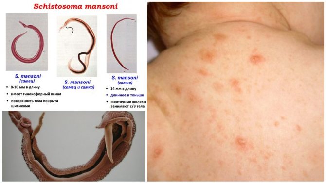
Schistosomiasis
Fans of traveling with children should be extremely careful when visiting these countries. Parasites enter the body through the skin when swimming in bodies of water. Male schistosomes reach 20 mm in size, females a little less than 15 mm, so it will not work to see how the worms look in the feces. Eggs of schistosomes are slightly oval, have no cap, but are equipped with a characteristic curved spine located along the body.
Schistosomiasis is a dangerous disease that causes mental and physical retardation in children. A child infected at an early age may look like a teenager at the age of 18.
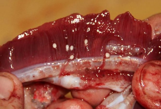
Pulmonary flukes
Are the cause of paragonimiasis in children. A worm up to 18 mm in size lives in the lungs of a child and has a body structure typical of this class with two suction cups, a uterus and testes. Outside, the body of the parasite is covered with small spines.
In the tissues of the lung, individuals are closed in a shell and mature for almost two years. Then they begin to lay eggs in a capsule next to them. Further, the eggs are released and, when coughing, can go out with phlegm into the mouth and from there be swallowed into the digestive tract. In this case, the eggs of these worms can be found in the child's feces and in the sputum. They are oval with a lid and are usually yellowish in color.
Infection of children with paragonimiasis occurs when eating crustaceans, which many boil only until reddening, which does not guarantee the death of pulmonary flukes.
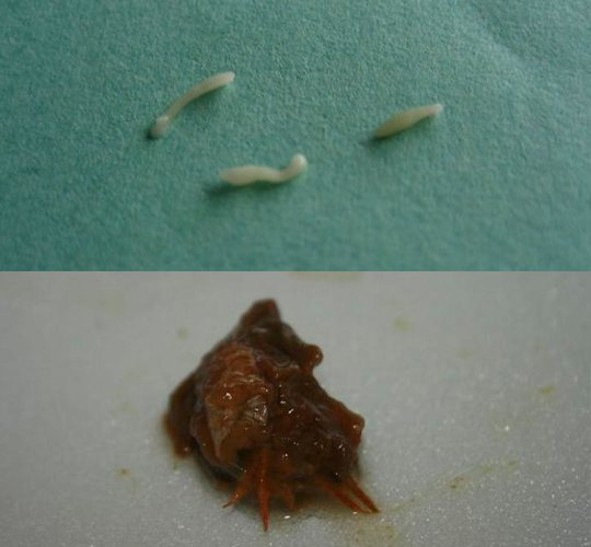
What do worms look like in feces in a child
Not everyone knows what helminths look like, so this is a hot topic. There are certain types of parasites that can be easily seen, others are very small, and special equipment is required to see such a worm.
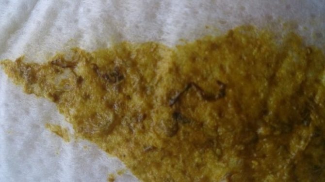
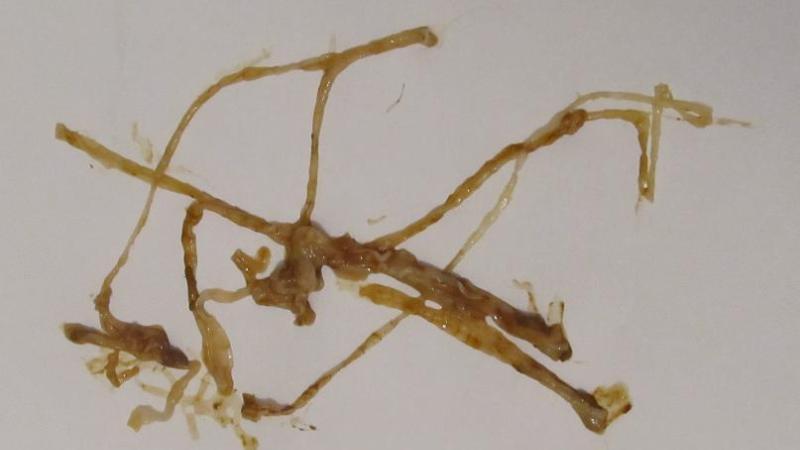
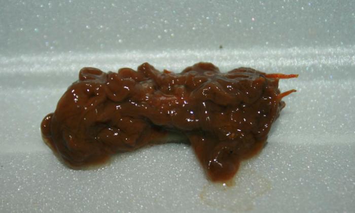
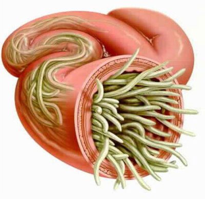
Sometimes the worms are excreted from the body in large tangles, in which there is a huge number of individuals.

If any specific inclusions appear, you need to contact a medical institution - only an experienced doctor can determine their nature, make an accurate diagnosis and prescribe adequate therapy.
Each worm is distinguished by its unique appearance and provokes certain diseases.
| Parasite | Disease | Appearance |
| Pinworms | Enterobiasis | Miniature worms of round size. They grow up to about 10 mm. |
| Roundworm | Ascariasis | They are long, roundworms, with a characteristic red and white hue, usually curved in a hook. Grow up to 6 cm. |
| Toksokara | Toxocariasis | Large specimen of round shape with a yellowish tinge. Reaches a length of about 10 cm. |
| Trichinella | Trichinosis | A small burgundy and round worm up to 5 mm long. |
| Vlasoglav | Trichocephalosis | It grows up to 5 cm. One end is thinner than the other. |
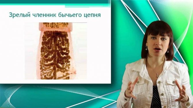
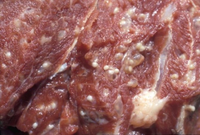
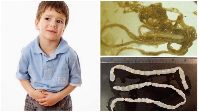
| Bovine tapeworm | Teniarinhoz | It is a long, ribbon-like worm with a small head and grows up to 30 m. |
| Pork tapeworm | Teniosis, Cysticercosis | Outwardly it looks like a bovine tapeworm, but of a shorter length - about 5 m. |
| Echinococcus | Echinococcosis | Small specimen - 11 mm. Differs in a tape shape, a head with suction cups, the body is divided into links. |
| Wide tapeworm | Diphyllobothriasis | The flat tapeworm reaches sizes up to 10 m. |
| Giardia | Giardiasis | Can be seen exclusively with a microscope. Some individuals are motionless, others move with the help of special flagella. |
| Feline or Siberian fluke | Opisthorchiasis | A small parasite no more than 10 mm long with a flat head. |
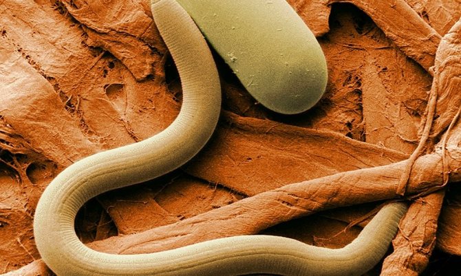
Roundworms or hookworms
The most common group of helminths in young children, which causes various types of nematodes.
A feature of round worms is the complete absence of the respiratory and circulatory systems. They absorb oxygen through the entire body.
The sizes of worms vary from small worms to medium, the habitat of individuals is various parts of the intestine. Pinworm is the causative agent of enterobiasis, which is often diagnosed in children under one year old.
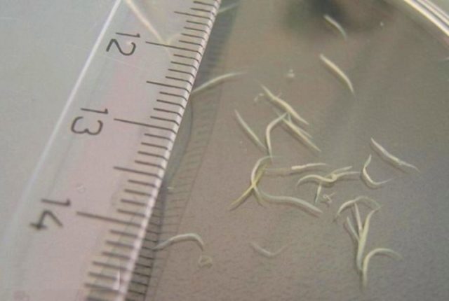
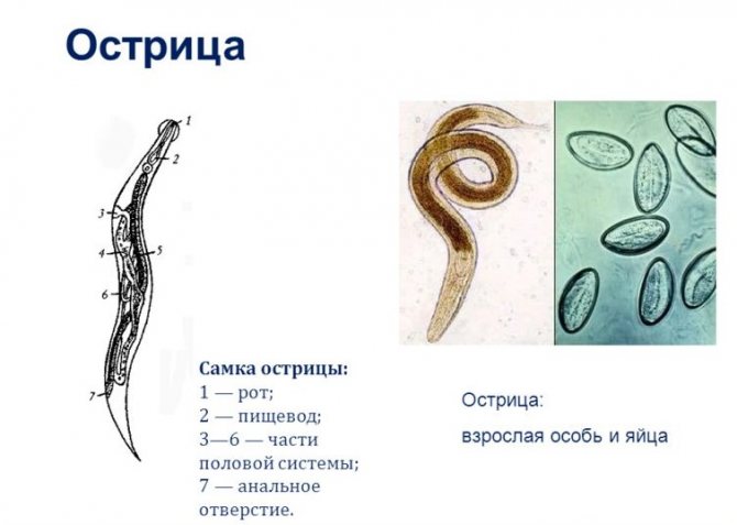
Pinworms
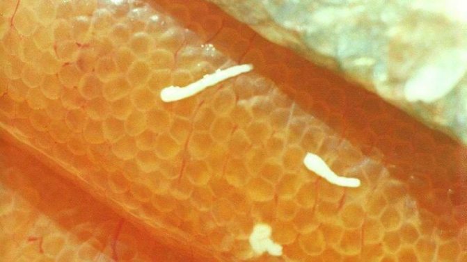
These are some of the most common worms in babies. They can be seen in a baby's pot with the naked eye.
- The sizes of females reach 12 mm, and males 5 mm. Their body shape is similar to a spindle.
- In females, the tip is pointed, in males it is twisted into a ring.
- Above, the body is slightly widened and equipped with a vesicle (mouth), with which the pinworm is attached to the intestinal wall. The oral cavity gradually merges into the esophagus, followed by the intestines and genital opening.
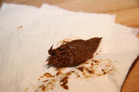
Pinworms have a special life cycle in which females die after laying eggs, and males after mating. Therefore, you can often see these parasites in the stool of a baby in large numbers. They look like scraps of white threads.
Many parents say they have seen similar black worms in their stool. But parasites do not have such a color. Most often, worms are taken for particles of undigested fiber from food.
Pinworm eggs are completely colorless. They are transparent, smooth, asymmetrical and multi-layered to help them survive in the environment.
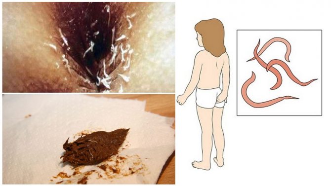
Pinworms in feces
Pinworms are the causative agent of enterobiasis. They can multiply en masse in babies' bodies and cause a number of pathological symptoms. These worms are often the cause of allergies and rashes in children.
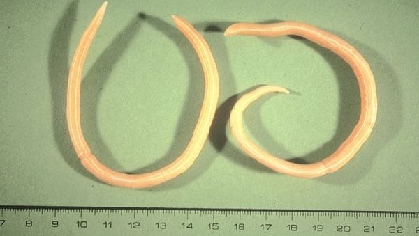
Roundworm
Many parents had to see this unsightly worm in a pot in a child. It is up to 20–40 cm long and never lives in the intestine in one specimen. To procreate, he needs a pair.
On a superficial examination, you might think that these worms are red, but in fact they are pinkish with a gray tint.
The male differs from the female in the lower end bent towards the abdomen. In the area of the head, roundworms have a mouth opening with three valves (lips). In the body of the worms there is a uterus, intestines and ovaries. On the cut, you can see the space between the organs (pseudo-goal). Ascaris eggs are elongated, small-knobby and filled with yolk-type cells for feeding the larvae.
Vlasoglav
This is far from a child's worm, but it often enters the child's intestines by the fecal-oral route and causes trichocephalosis. Vlasoglav has a special structure, for which it got its name. The head of the worm is like a hair, it contains a mouth and a long esophagus, through which the individual sucks nutrients from the intestinal membranes. The size of the parasite is on average 5 cm. The reproductive system and intestines are located in the posterior expanded part of the whipworm.
Larval worm capsules look like small barrels with a thick shell. They are transparent, with caps on two poles and filled with the contents of a fine-grained structure.
In rare cases, children may develop other roundworms. These are toxocars, trichinella and hookworms. They have a similar structure and also enter the body through the mouth.
Is it possible to see the eggs of worms in the feces of a child
Drits Irina Alexandrovna. Parasitologist Helminthiasis can lead to numerous health problems, shortening life by 15-25 years. Many parasites are extremely difficult to detect. They can be anywhere - in the blood, intestines, lungs, heart, brain. Symptoms of helminthic invasion can be confused with ARVI, gastrointestinal diseases and others. The main mistake in such cases is procrastination! If you have any suspicions about the presence of parasites, then you need to see a specialist. More information about modern methods of treating helminthiasis is described in this interview with a doctor... If we talk about drugs and self-treatment, then from the most common helminths (ascaris, pinworms, tapeworms), this antiparasitic complex.
It is extremely difficult to see the eggs of the parasite in the feces, since they differ in microscopic size. Parents can find streaks in the baby's feces in the form of thin threads of white, black or red.
Usually, dark streaks appear after eating bananas, white indicates the presence of parasites, and red indicates blood.
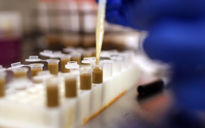

Diagnostics
To identify parasitic infection, the patient must pass the appropriate tests. Diagnosis is by microscopy of the perianal scraping. An analysis for enterobiasis should be done in the morning before bowel movements and before hygiene procedures. It is carried out in two ways: using adhesive tape (attached to the anus) and a cotton swab with petroleum jelly.
Such a diagnosis can give false negative results, since female pinworms do not lay eggs every day. For this, microscopy is performed 2-3 times every two weeks.
At the same time, the usual analysis of feces for pinworm eggs is not informative. This is due to the fact that eggs are not deposited in the intestines, but near the anus. Also, the patient is assigned a general blood test. With parasitic invasion, the level of eosinophils is increased. This indicates an allergic reaction of the body to the introduction of helminths. If an inflammatory reaction is observed, then this indicates a complicated course of invasion from the gastrointestinal tract.
[], []
Perianal scraping for pinworm eggs
The procedure for taking biomaterial around the anus to diagnose enterobiasis is a perianal scraping of pinworm eggs. This analysis allows you to establish the presence of parasites with an accuracy of 90%. Before passing it, you need to do a little preparation in order to get reliable results:
- Do not go to the toilet before scraping.
- Do not wash off as you can wash off all pinworm eggs.
- Do not take a laxative the day before the procedure.
Scraping is done with a cotton swab, duct tape or wooden spatula. If the procedure is performed at home and a cotton swab is used, then after analysis it should be placed in an airtight, clean container. The adhesive tape must be delivered to the laboratory for testing within 8 hours after the material has been taken.
Perianal scraping is taken very quickly.You can get the results of the analysis every other day, since the material is valid for 24 hours. If the result is negative, but there are symptoms of invasion, then the verification algorithm is repeated after 2-3 days. It should also be noted that the diagnostic result is valid for 10 days. After their expiration, the analysis must be repeated.
Does feces change with worms
The structure of feces with helminthic invasion can change at the stage of exacerbation, when the child develops any form of stool disorder. Feces can be liquid, ordinary.
With prolonged constipation, it is better to resort to the use of an enema, which will help thin the feces so that they leave the intestines without damaging its mucous membrane.
Prophylaxis
Any disease is better prevented than cured. Moreover, this applies to antihelminthic drugs. Prevention of helminthic invasion plays a huge role in human life.
Observe the following rules:
- keep your child's hands clean;
- wash fruits and vegetables thoroughly;
- heat meat and fish products;
- with constant contact with animals, use antihelminthic drugs in a prophylactic dosage.
Loading ...
White worms in a child in feces
White worms in feces indicate pinworm infestation. This is the most common type of parasite that infects the human body.
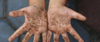
Traditionally, the cause of infection is a violation of hygiene rules. Worms enter the body through unwashed hands, food and settle in the intestines, sometimes in other organs.
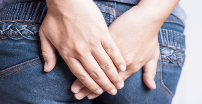
The female is able to lay approximately 15,000 eggs in the intestines and around the anus, which mature within 6 hours. During this process, the baby feels severe itching, especially in the morning and evening (the period of activity of the parasite).
Since the child will scratch the problem area, the eggs fall under his nails, on underwear and other objects, which contributes to their spread.
After mating, the males die and go out with the feces. In the feces, you can see both living and dead individuals of both sexes.
Diagnostics

To make a diagnosis, it is not enough to rely only on data from an external examination. It is imperative to carry out instrumental and laboratory research methods. To establish the presence of helminths in the body, you need to take material for research, where you can find adults, eggs, larvae. Usually it is feces, bile, urine, blood, sputum, a piece of muscle tissue. Any of these materials are specially processed and viewed under a microscope. The most commonly used analysis is the delivery of feces for helminth eggs. But this method is not always informative, since the eggs of not all worms are found in the human intestine (for example, with enterobiasis).
A blood test for all helminthiases shows eosinophilia of varying severity. With massive invasion, severe or prolonged course of the disease, changes such as a decrease in the level of red blood cells and hemoglobin may appear.
In a biochemical blood test, a decrease in the level of total protein and the amount of albumin is possible, especially in the case of infection with trichinosis. Liver tests (thymol test, total bilirubin, alkaline phosphatase level) can change when infected with opisthorchiasis or fascioliasis. Changes in the analysis of urine will be with schistosomiasis (hematuria, leukocyturia, cylindruria).
Serological research methods are widely used. To detect helminth antigens and antibodies formed against them, enzyme immunoassay, indirect and passive hemagglutination reaction, immunofluorescence reaction, and complement binding reaction are used. The most informative method is PCR (polymerase chain reaction), which finds the pathogen by the structure of its DNA or RNA.
Instrumental research methods are used to check the state of internal organs. All persons with suspected helminthiasis and with dysfunction of the digestive canal are assigned an ultrasound scan of the abdominal organs, hepatobiliary system. With schistosomiasis, ultrasound of the bladder and kidneys can be done.
With ascariasis, X-ray is necessary to detect infiltrates in the lungs. With trichinosis, a muscle biopsy study will be important. In all cases, CT (computed tomography) can be used as an additional examination, which will more accurately indicate the localization of pathological foci, the degree of organ damage, and will make it possible to determine further therapy.
Small worms in the feces of a child
If you find small worms in the stool of a child, you need to contact a medical institution. After passing the tests, the doctor will make an accurate diagnosis and select adequate methods of therapy.
Take a worm test
The presence of worms in the feces indicates a helminthic invasion, one of the many types of parasites. It is strictly forbidden to leave such a problem unattended - the advanced stage of infection will lead to a serious negative effect on the child's body.
First signs
The acute stage of helminthiasis begins two weeks after infection. Signs of worms in humans differ depending on the type of pest. The first symptoms of the disease:
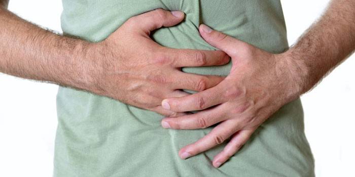
- stomach ache;
- lack of appetite;
- fever;
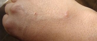
- rashes on the skin;
- airway inflammation;
- conjunctivitis;
- loose stools;
- drastic weight loss;
- itching in the anus.
When pests are localized in the intestines, they produce substances similar to hormones that cause disturbances in the functions of the gastrointestinal tract. This process is characterized by signs:
- prolonged diarrhea;
- flatulence;
- severe pain in the right hypochondrium, near the navel;
- intestinal obstruction - with a large number of individuals;
- chronic constipation;
- nausea;
- periodic vomiting.
The appearance of worms causes symptoms associated with body poisoning:
- Nervous system changes - mood swings, headache, vomiting, nightmares, insomnia, distraction. Children have problems with studies, whims.
- Allergic reactions - dry cough, rhinitis, hives, skin rashes.
- Decreased immunity, provoking the development of infectious diseases, exacerbation of chronic pathologies, gynecological inflammation.
- The appearance of helminths in the feces.
- Temperature increase.
- Brittle nails, cracked heels, hair loss.
- Grinding of teeth.
What to do and how to treat
If a child is diagnosed with infection with worms, the doctor selects the means of therapy. The treatment is comprehensive, first you need to prepare the body with a diet, then antibacterial agents are used.
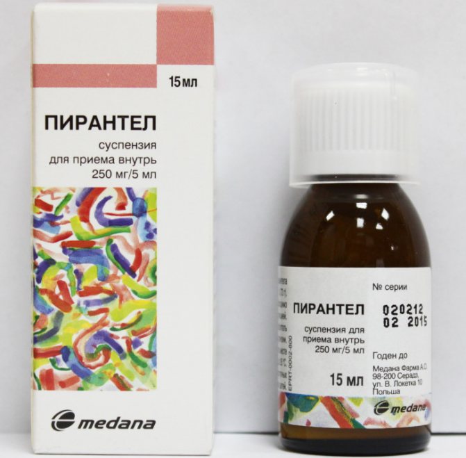
To restore the body, vitamin complexes and preparations are prescribed to strengthen the immune system. Usually for the treatment of helminths are prescribed:
- Pirantel;
- Helmintox;
- Mebendazole;
- Vermox;
- Nourished.

It is categorically not recommended to choose medicines on your own - all antihelminthic drugs are toxic, have many contraindications and side effects. They can be prescribed exclusively by the attending physician.
In addition to traditional methods of therapy, folk remedies show high efficiency. However, before using any prescription, you need to consult with your doctor, as they also have certain contraindications and can aggravate the problem.
During the preparation of a recipe, it is important to observe the dosage indicated in it, otherwise side effects may develop!
Among the popular folk remedies are:
- pumpkin seeds;
- garlic;
- infusion of pomegranate peel;
- carrot juice;
- vegetable oils;
- decoctions of tansy and wormwood.
It is important for parents not only to monitor the well-being of the child, but also to check his feces for the presence of parasites. This is the best way to identify the invasion in time and provide the baby with timely medical care.
It is impossible to ignore the infection - the process of vital activity of worms negatively affects the functioning of many internal organs, systems and health in general.
You can defeat parasites!
Antiparasitic complex® - Reliable and safe disposal of parasites in 21 days!
- The composition includes only natural ingredients;
- Does not cause side effects;
- Absolutely safe;
- Protects liver, heart, lungs, stomach, skin from parasites;
- It removes the waste products of parasites from the body.
- Effectively destroys most types of helminths in 21 days.
There is a preferential program now for free packaging. Read expert opinion.


Factors affecting the manifestations of helminthiasis
The way the parasite enters the body;- The degree of adaptation of the helminth to the human body;
- Population density (number) of parasitic individuals;
- The habitat of the worm (tissue parasites live in the thickness of soft tissues, and luminal ones live in the gaps of hollow organs). Some helminths in different phases have both luminal and tissue forms. Larval and developing stages of worms, as a rule, cause more pronounced pathological changes.
In the absence of re-infection, the number of adult parasites in the human body does not increase. This feature significantly distinguishes helminthic invasions from diseases caused by bacteria, viruses, fungi and protozoa.